Author: admin

  • كل ما يلزمك معرفته حول نقص اليود عند الأطفال

    الكاتب: ميس حسن /الدليل-الطبي/%D9%85%D9%8A%D8%B3-%D8%AD%D8%B3%D9%86-192736 | تاريخ النشر: تاريخ التعديل 11 نوفمبر 2024 | 7 دقيقة قراءة

    المصدر الأصلي: الطبي – Altibbi

    كل ما يلزمك معرفته حول نقص اليود عند الأطفال

    المشاركة عبر وسائل التواصل الاجتماعي

    يُمكن أن يُؤثر نقص اليود عند الأطفال سلبًا على صحتهم، وربما يُعرضهم لمشكلاتٍ خطيرة تؤثر على نموهم، ورغم أن الأطفال يحتاجون اليود بكميات قليلة، إلا أنه يجب التأكد من حصول الطفل على الكمية الموصى بها من اليود، والتي تختلف باختلاف مراحل نموه. [7]

    ما هو نقص اليود عند الأطفال؟ وما هي أسبابه؟ وكيف يمكن علاجه والوقاية منه؟ إليك التفاصيل في المقال الآتي.

    محتويات المقال

    نقص اليود عند الأطفال

    يحدث نقص اليود عند الأطفال عندما لا يحصل الجسم على حاجته من اليود، [3] ويُعد اليود من العناصر الأساسية لنمو الأطفال؛ لدوره في إنتاج هرمونات الغدة الدرقية (بالإنجليزية: Thyroid Hormones) بنوعيها: ثلاثي يودوثيرونين (T3) وثيروكسين (T4)، وهي الهرمونات المسؤولة عن تنظيم عملية الأيض أو التمثيل الغذائي، ومعدل ضربات القلب، والعديد من الوظائف الحيوية الأخرى، بالإضافة إلى دورها الهام في نمو الدماغ، والأعصاب، والعظام. [6][7]

    وعادةً يحصل الجسم على حاجته من اليود عن طريق الطعام، خاصةً المأكولات البحرية وملح الطعام المعالج باليود، لذا فإن نقص اليود عند الأطفال يكون أكثر شيوعًا لدى الأطفال حديثي الولادة؛ بسبب اعتمادهم على الرضاعة، والأطفال الذين لا يتناولون كمية كافية من الأطعمة الغنية باليود. [3]

    اقرًا أيضًا: فوائد اليود المحتملة

    مستوى اليود المنخفض عند الأطفال

    يُفرز الجسم اليود الذي يتناوله الطفل عن طريق البول، لذا غالبًا يعتمد الأطباء على نسبة اليود في البول لتقييم مستواه في الجسم، وبحسب توصيات منظمة الصحة العالمية فإن كمية اليود التي يتناولها الطفل تكون كافية إذا كان معدل اليود في البول يتراوح بين 100- 199 ملليجرام/لتر. [3]

    في حين يدل انخفاض نسبة اليود في البول عن هذا المعدل على نقص اليود عند الطفل، وفيما يأتي تفصيل ذلك: [3]

    • يكون الطفل مصابًا بنقص اليود الشديد إذا كان معدل اليود في البول أقل من 20 ملليجرام/لتر.
    • يكون الطفل مُصابًا بنقص اليود المتوسط إذا كان معدل اليود في البول بين 20-49 ملليجرام/لتر.
    • يكون الطفل مُصابًا بنقص اليود الخفيف إذا كان معدل اليود في البول بين 50-99 ملليجرام/لتر.

    هل لديك اسئلة متعلقة في هذا الموضوع؟
    اسال سينا، ذكاء اصطناعي للاجابة عن كل اسئلتك الطبية
    اكتب سؤالك هنا، سينا يجهز الاجابة لك

    أسباب وعوامل خطر نقص اليود عند الأطفال

    يحدث نقص اليود عند الأطفال بسبب عدة عوامل، أهمها:[5][6][8]

    • عدم حصول الأم على كميات كافية من اليود خلال الحمل أو الرضاعة، مما يسبب نقص اليود عند الجنين أو الرضيع.
    • عدم حصول الطفل على اليود بشكل كافٍ من خلال الغذاء، خاصة عند بدء الطفل بتناول الأطعمة الصلبة.
    • احتواء التربة على كميات قليلة من اليود، مما يؤدي إلى انخفاض نسبة اليود في المزروعات من الخضروات والفواكه.
    • حاجة الطفل لاتباع حمية تُركز على تناول كمياتٍ قليلة من الملح، كما هو الحال مع الأطفال المصابين بالتوحد.
    • انتقائية الطفل في تناول الطعام، أو بمعنىً آخر تركيزه على تناول أنواعٍ معينة فقط من الأطعمة مثل: الأرز، والموز، وحبوب الإفطار، مما يجعله أكثر عرضة للإصابة بنقص اليود والمعادن بشكلٍ عام.

    أعراض نقص اليود عند الأطفال

    يرتبط نقص اليود غالبًا بانخفاض هرمونات الغدة الدرقية، مما يُؤدي إلى الإصابة بقصور أو كسل الغدة الدرقية (بالإنجليزية: Hypothyroidism) في حال استمر نقص اليود لدى الطفل لسنوات أو كان شديدًا. [5]

    ويُؤدي إصابة الطفل بكسل الغدة الدرقية إلى ظهور مجموعة من الأعراض، أبرزها: [2][7][8]

    • تضخم الغدة الدرقية (بالإنجليزية: Goiter)، هو من العلامات الشائعة لنقص اليود، ويظهر على هيئة تضخم في الجزء الأمامي من الرقبة، وذلك كمحاولة من الغدة الدرقية لامتصاص كمياتٍ أكبر من اليود الموجود في الدم، وأحيانًا يسبب التضخم الشديد صعوبة في التنفس والبلع.
    • زيادة الوزن، بسبب نقص هرمونات الغدة الدرقية، والذي يُبطئ عملية الأيض وحرق السعرات الحرارية، ويسبب تخزين السعرات الحرارية على شكل دهون.
    • تساقط الشعر، خاصةً أنّ هرمونات الغدة الدرقية تنظم عملية نمو بصيلات الشعر، ولكن في المقابل فإن الشعر يستعيد نموه الطبيعي عند علاج مشكلة نقص اليود وقصور الغدة.
    • التعب والخمول، حيث تُساعد هرمونات الغدة الدرقية على تنظيم الطاقة، لذا فإن نقص اليود وانخفاض هذه الهرمونات يُؤدي لشعور الطفل بالتعب الزائد وقلة النشاط.
    • عدم القدرة على تحمل البرودة، وذلك بسبب بطء عملية الأيض وانخفاض الطاقة، مما يُؤدي لشعور الطفل بالبرد الزائد.
    • جفاف الجلد، خاصةً أنّ انخفاض هرمونات الغدة يُسبب قلة التعرق، ويجعل عملية تجديد خلايا الجلد أبطء.
    • مشكلات في الذاكرة والتعلم، إذ لوحظ أن منطقة الدماغ المسؤولة عن الذاكرة يكون حجمها أصغر عند المصابين بقصور الغدة الدرقية الناجم عن نقص اليود.
    • أعراض أخرى، مثل: الإمساك، وبطء ضربات القلب خاصةً إذا كان نقص اليود شديدًا.

    تشخيص نقص اليود عند الأطفال

    يُعتمد الطبيب على عددٍ من الفحوصات لتشخيص الطفل بنقص اليود، أهمها: [2][3][4]

    • فحص تركيز اليود في البول، والذي يقيس معدل تناول الطفل لليود خلال الأيام السابقة.
    • فحص الدم للكشف عن مستويات الهرمون المنبه للغدة الدرقية (Thyroid Stimulating Hormone)، وهرمون T3، وT4؛ خاصةً إذا كان نقص اليود شديدًا أو ظهر على الطفل علامات تدل على إصابته بكسل الغدة الدرقية.
    • فحص بروتين الغلوبيولين الدرقي (بالإنجليزية: Thyroglobulin)، يُساعد هذا الفحص على تشخيص قصور الغدة الدرقية وفرط نشاطها أيضًا، كما يستخدمه الطبيب لتقييم مستويات اليود لدى الطفل خلال الأسابيع والأشهر الماضية.
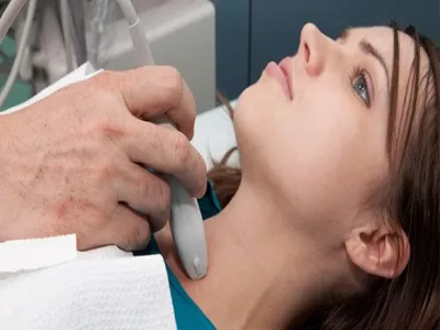
    • تصوير الغدة الدرقية بالموجات فوق الصوتية إذا كان الطفل مصابًا بتضخم الغدة الدرقية.

    sina inread banner image
    مساعدك الشخصي من الطبي للاجابة على أسئلتك الصحية
    الطبي يطلق سينا، ذكاء اصطناعي لخدمتك الصحية!
    اسأل سينا

    علاج نقص اليود عند الأطفال

    يلجأ الطبيب إلى الطرق الآتية لعلاج نقص اليود: [2][5]

    • استعمال الملح المعالج باليود أثناء تحضير الطعام، والتي تُعد من أفضل طرق العلاج وأقلها تكلفة.
    • تناول الأطعمة الغنية باليود، مثل:
      • الخبز المدعم باليود.
      • المأكولات والأعشاب البحرية.
      • البيض ومنتجات الألبان.
      • الفواكه والخضروات.
      • اللحوم.
    • تناول المُكملات الغذائية التي تحتوي على اليود، وذلك بعد استشارة الطبيب أولًا.
    • الخبز المدعم باليود.
    • المأكولات والأعشاب البحرية.
    • البيض ومنتجات الألبان.
    • الفواكه والخضروات.
    • اللحوم.

    طرق الوقاية من نقص اليود عند الأطفال

    يُمكن الوقاية من نقص اليود ومنع المضاعفات التي يُسببها بالالتزام بالجرعات اليومية الموصى بها من اليود، والتي تختلف باختلاف عمر الطفل، ويُوضح الجدول الآتي الجرعات الموصى بها من اليود للأطفال: [7][8]

    الجرعة الموصى بها من اليود يوميًا (ميكروغرام)

    ومن الضروري أن تحصل الأم المرضعة على 290 ميكروغرام يوميًا من اليود؛ لضمان حصول رضيعها على كمية كافية من اليود، أمّا بالنسبة للجرعة الموصى بها يوميًا للأم الحامل فهي تبلغ 220 ميكروغرام.[7]

    ويُمكن الحصول على الكميات السابقة الموصى بها من اليود بإحدى الطرق الآتية: [2][7]

    • يُفضل أن تتناول الأم الحامل أو المرضع المكملات الغذائية التي تحتوي 150 ميكروغرام من اليود يوميًا على الأقل، لضمان حصول الجنين أو الرضيع على كميات كافية منه؛ لأنّ آثار نقص اليود تكون أكثر خطورة عند الأجنة والرضع.
    • استعمال الملح المعالج باليود لتحضير الطعام، خاصةً إذا كان الطفل يرفض تناول الخبز، أو منتجات الألبان، وتحتوي كل ملعقة طعام من هذا الملح على 250 ميكروغرام تقريبًا من اليود.
    • تناول الأطعمة التي تحتوي اليود.

    للمزيد: أبرز الأطعمة الغنية باليود

    مضاعفات نقص اليود عند الأطفال

    يُسبب نقص اليود مضاعفات قد تكون خطيرة أحيانًا لدى الأطفال أو الرضع،[1] أهمها: [5][6][7]

    • تأخر أو إعاقة ذهنية لدى الرضع والأطفال الذين لم تحصل أمهاتهم على كمية كافية من اليود خلال فترة الحمل، لدوره في عملية تطور دماغ الجنين ونموه.
    • انخفاض وزن الجنين عند الولادة، ونتيجةً لذلك ربما يحتاج الطفل لتلقي العلاج في الحضانة (الخداج).
    • زيادة خطر ولادة جنين ميت.
    • تلف الدماغ عند الرضع أو الأطفال الأصغر سنًا، مما يُؤثر على نموهم العقلي والحركي وأدائهم الدراسي مستقبلًا.
    • قصور الغدة الدرقية (بالإنجليزية: Hypothyroidism) أو تضخمها، لذلك يحتاج الطفل إلى اليود بشكل أساسي خلال أول عامين من حياته.
    • تأخر نمو الطفل.

    يرتبط نقص اليود عند الأطفال بمشكلات صحية عديدة، مثل قصور عمل الغدة الدرقية، والتي يحتاج الطفل إلى هرموناتها في نمو أجهزة جسمه ودعم وظائفه الحيوية. في المقابل يمكن علاج النقص بتناول الملح المُعالج باليود، والأطعمة الغنية باليود، والمكملات الغذائية حسب توصيات الطبيب.

    ويُمكنك الآن الحصول على استشارة منزلية في أي وقت ومن أيّ مكان بمساعدة أحد أطبائنا المعتمدين على منصة الطبي.

    اقرا ايضاً :

    أربع فحوصات لتقييم العقد الدرقية

    اقرا ايضاً :

    الاختلال بافراز الغدة الدرقية فرط النشاط او القصور اصبح امرا شائعا ولا يدعو للقلق ولكن ما يثير المخاوف حاليا هو … اقرأ أكثر

    [1] World Health Organisation (WHO). Nutrition: Effects of iodine deficiency. Retrieved on the 28th of September, 2024.

    [2] Health direct. Iodine deficiency. Retrieved on the 28th of September, 2024.

    [3] Caring for Kids New to Canada. Iodine Deficiency. Retrieved on the 28th of September, 2024.

    [4] Medline Plus. Thyroglobulin. Retrieved on the 28th of September, 2024.

    [5] Raising Children.net.au. Iodine: what you need to know. Retrieved on the 28th of September, 2024.

    [6] Michigan Medicine. Pass the Salt: Iodine Crucial for Kids’ Diets. Retrieved on the 28th of September, 2024.

    [7] Dr. Sravani Chintapalli. 10 Signs and Symptoms of Iodine Deficiency. Retrieved on the 28th of September, 2024.

    [8] Paul Frysh. Are You Getting Enough Iodine? Retrieved on the 28th of September, 2024.

    المصدر: الطبي – Altibbi

    هذا المقال مقدم لأغراض إعلامية فقط وليس بديلاً عن المشورة الطبية المهنية. يُرجى استشارة مقدم رعاية صحية مؤهل للحصول على التشخيص والعلاج.

  • علاقة الغدة الدرقية ورائحة الفم الكريهة

    الكاتب: رغد زعبلاوي /الدليل-الطبي/%D8%B1%D8%BA%D8%AF-%D8%B2%D8%B9%D8%A8%D9%84%D8%A7%D9%88%D9%8A-159810 | تاريخ النشر: تاريخ التعديل 21 سبتمبر 2024 | 6 دقيقة قراءة

    المصدر الأصلي: الطبي – Altibbi

    علاقة الغدة الدرقية ورائحة الفم الكريهة

    المشاركة عبر وسائل التواصل الاجتماعي

    تُعدّ الغدة الدرقية إحدى الغدد المهمة مهم جدًا في جسم الإنسان، فهي تلعب دورًا رئيسيًا في عملية التمثيل الغذائي، وبالتالي فهي تؤثر على وظائف الأعضاء بشكل كبير. ومن المُمكن أن تؤثر اضطرابات الغدة الدرقية على الصحة العامة لمختلف أعضاء الجسم، بما في ذلك الفم، ومن المُمكن أن تكون هذه الاضطرابات هي السبّب الرئيسي وراء انبعاث رائحة كريهة من الفم. [1]

    وفي هذا المقال سنتعرف على العلاقة ما بين الغدة الدرقية ورائحة الفم، وكيف يُمكن التخلص من هذه المشكلة.

    محتويات المقال

    علاقة الغدة الدرقية ورائحة الفم

    تنتج رائحة الفم الكريهة بشكل رئيسي عن إطلاق مركبات الكبريت المتطايرة، والتي تنتج بسبب تراكم البكتيريا في الفم. ويزداد إنتاج هذه المركبات لدى الأشخاص الذين يعانون من سوء نظافة الفم ومشاكل اللثة والفم، كما يُمكن أن تزداد أيضًا لدى أولئك الذين يعانون من اضطرابات الغدة الدرقية والأمراض المزمنة الأخرى. [1]

    ومن الجدير ذكره أنّ رائحة الفم الكريهة لا ترتبط ارتباطًا مباشرًا باضطرابات الغدة الدرقية، بل يُمكن أن تكون نتيجةً لتأثير هذه الاضطرابات على وظائف الجسم المختلفة. فعلى سبيل المثال، يمكن أن تؤدي اضطرابات الغدة الدرقية إلى جفاف الفم واضطرابات في الجهاز الهضمي، وقد يترتب على ذلك زيادة احتمالية الإصابة برائحة الفم الكريهة. [1][2]

    بالإضافة إلى ذلك، إنّ نشاط الغدة الدرقية يُمكن أن يرتبط بمتلازمة شوغرين، وهو ما يُمكن أن يؤثر على إفراز اللعاب، وبالتالي يزيد من احتمالية حدوث رائحة الفم الكريهة. [1][2]

    وفيما يلي نذكر الأسباب الكامنة وراء العلاقة ما بين أمراض الغدة الدرقية ورائحة الفم الكريهة بالتفصيل:

    جفاف الفم

    من بين المضاعفات التي يُمكن أن تنتج عن أمراض الغدة الدرقية وتؤدي إلى رائحة الفم الكريهة، يأتي انخفاض إنتاج وإفراز اللعاب من الغدد اللعابية في المقدمة، والذي غالبًا ما يُسبّب جفاف الفم. [1][2]

    ويعود السبّب في هذا الانخفاض في إنتاج اللعاب لبطء عملية التمثيل الغذائي المرتبطة بقصور الغدة الدرقية. بالإضافة إلى تسبب قصور الغدة الدرقية في التنفّس عبر الفم أثناء النوم والشخير، وهي إحدى الأسباب الشائعة جدًا لجفاف الفم ورائحة الفم الكريهة. [1][2]

    وفي حين أن جفاف الفم يرتبط غالبًا بقصور الغدة الدرقية، إلا أنّه من المُمكن أن يحدث أيضًا مع مجموعة أخرى من أمراض الغدة الدرقية، والتي تشمل: [2]

    • فرط نشاط الغدة الدرقية غير المعالج أو غير المسيّطر عليه، ولكن السبّب وراء هذا الارتباط غير واضح حتى الآن ويحتاج إلى المزيد من البحث والدراسة.
    • التهاب الغدة الدرقية لهاشيموتو (بالإنجليزية: Hashimoto’s Thyroiditis)، وهو أحد أنواع أمراض المناعة الذاتية التي تُسبّب قصور في وظائف الغدة الدرقية.
    • التهاب الغدة الدرقية تحت الحاد (بالإنجليزية: Subacute Thyroiditis)، والذي قد ينجم عن أسباب مختلفة، مثل: الإصابة بأنواع أخرى من التهاب الغدة الدرقية، أو استخدام بعض الأدوية، أو الإصابة بعدوى، ويُمكن أن تؤدي هذه الحالة إلى تطوّر قصور الغدة الدرقية أو فرط نشاطها.
    • قصور الغدة الدرقية غير المشخّص (بالإنجليزية: Subclinical Hypothyroidism)، حيث يُمكن أن يعاني الأشخاص المصابون بقصور الغدة الدرقية في مراحل مبكرة من أعراض غير ملحوظة جدًا، بما في ذلك جفاف الفم.
    • مرض جريفز (بالإنجليزية: Grave’s Disease)، وهو نوع من أنواع فرط نشاط الغدة الدرقية الشائعة، والذي يُسبب أعراضًا مثل: فقدان الوزن، والأرق، وجفاف الفم.

    وإلى جانب الأمراض التي تصيب الغدة الدرقية، تُعدّ متلازمة شوغرن (بالإنجليزية: Sjogren’s Syndrome) واحدة من المتلازمات الأكثر شيوعًا لدى الأشخاص المصابين بالتهاب الغدة الدرقية هاشيموتو والتي تُسبّب جفاف الفم ورائحة الفم الكريهة. وفي الوقت نفسه، يُعتبر مرضى متلازمة شوغرن أكثر عرضة لخطر الإصابة بمشاكل في الغدة الدرقية. [1][3]

    متلازمة شوغرن هي إحدى الأمراض المناعية الذاتية التي تؤثر بشكل أساسي على الغدد التي تنتج الدموع واللعاب، مما يؤدي إلى جفاف العين والفم. ويعتبر هذا النقص في إنتاج اللعاب سببًا رئيسيًا يجعل الفم بيئة خصبة لنمو البكتيريا، مما يؤدي إلى انبعاث رائحة كريهة من الفم. [1]

    ولا تقتصر العلاقة بين اضطرابات الغدة الدرقية ورائحة الفم الكريهة الناتجة عن جفاف الفم بالأمراض بحد ذاتها، ولكن يُمكن أن تُسبّب بعض علاجات أمراض الغدة الدرقية جفاف الفم لدى المريض، ومن بينها: [2]

    • العلاج الإشعاعي: حيث يُمكن أن يؤثر العلاج الإشعاعي المستخدم في علاج سرطان الغدة الدرقية على الغدة الدرقية، الأمر الذي قد يؤدي إلى تلف الغدد اللعابية، ممّا يقلل من كمية اللعاب التي يتم إنتاجها في الفم.
    • العلاج باليود المشع: فمن المُمكن أن يؤدي استخدام اليود المشع إلى قصور الغدة الدرقية وانخفاض كمية اللعاب المفرزة من قبل الغدة اللعابية. ومن الجدير بالذكر أن اليود المشع يستخدم لعلاج سرطان الغدة الدرقية وبعض أنواع فرط نشاط الغدة الدرقية.
    • العلاج الهرموني: فعلى الرغم من أن استخدام العلاج البديل لهرمون الغدة الدرقية يساعد في تحسين جفاف الفم، إلا أنّه في بعض الحالات قد يؤدي إلى جفاف الفم.

    أمراض الفم واللثة

    تُسبّب اضطرابات الغدة الدرقية غير المسيطر عليها زيادة خطر الإصابة بالعديد من الأمراض التي تؤثر على الفم واللثة، والتي تُعدّ من أبرز أسباب رائحة الفم الكريهة. وتشمل اضطرابات الفم الناتجة عن اضطرابات الغدة الدرقية ما يلي: [3][4]

    • ضعف صحة اللثة.
    • تأخر التئام الجروح في الفم.
    • تسوس الأسنان.
    • هشاشة الفك العلوي والسفلي.
    • متلازمة الفم الحارق.

    sina inread banner image
    مساعدك الشخصي من الطبي للاجابة على أسئلتك الصحية
    الطبي يطلق سينا، ذكاء اصطناعي لخدمتك الصحية!
    اسأل سينا

    اضطرابات الجهاز الهضمي

    ترتبط اضطرابات الغدة الدرقية مع رائحة الفم الكريهة أيضًا نتيجةً لتأثيرها على صحة الجهاز الهضمي، حيث تؤدي اضطرابات الغدة الدرقية، وعلى وجه الخصوص قصور الغدة الدرقية، إلى إبطاء عملية التمثيل الغذائي، ما يُسبّب إبطاء حركة الطعام في الجهاز الهضمي، والذي غالبًا ما يؤدي إلى مشاكل في الهضم، والإصابة بالإمساك، والارتجاع المريئي. [1]

    ويُعدّ الارتجاع المريئي من الأسباب الشائعة للإصابة برائحة الفم الكريهة لسببين، وهما: [1][4]

    • يحدث ارتجاع المريء عندما تتدفق محتويات المعدة، مثل: حمض المعدة وبقايا الطعام غير المهضوم، بشكل عكسي إلى المريء، مسببًا رائحة الفم الكريهة.
    • يؤدي ارتجاع المريء إلى تآكل طبقة مينا الأسنان، ممّا يزيد من خطر الإصابة بتسوس الأسنان والتجاويف، والتي تُعدّ أيضًا من أسباب رائحة الفم الكريهة.

    علاج رائحة الفم الناجمة عن الغدة الدرقية

    تُعدّ الخطوة الأولى في علاج رائحة الفم الكريهة هي تحديد السبّب. وعندما تكون اضطرابات الغدة الدرقية هي سبب هذه المشكلة، غالبًا ما يقوم الطبيب بوصف دواء يعمل على تعديل مستويات هرمون الغدة الدرقية في الجسم، ممّا يساعد وظائف الجسم بالعودة إلى وضعها الطبيعي، وبالتالي سيتم علاج رائحة الفم الكريهة المرتبطة بالغدة الدرقية. [1][5]

    وتشمل طرق علاج اضطرابات الغدة الدرقية الشائعة ما يلي: [5]

    • في حال الإصابة بقصور الغدة الدرقية، سيصف الطبيب دواء بديل لهرمون الغدة الدرقية، ويُعرف باسم الليفوثيروكسين (بالإنجليزية: Levothyroxin).
    • في حال الإصابة بفرط نشاط الغدة الدرقية، فيتم علاجها باستخدام اليود المشع، والأدوية المضادة للغدة الدرقية، وقد يلجأ الطبيب إلى الجراحة.

    وإلى جانب استخدام العلاج المناسب لاضطرابات الغدة الدرقية، يُعدّ الحفاظ على نظافة الفم أمرًا ضروريًا للتخلص من رائحة الفم الكريهة، وهذا يتضمّن ما يلي: [1][4]

    • تنظيف الأسنان مرتين يوميًا لمدة دقيقتين على الأقل، باستخدام معجون أسنان يحتوي على الفلورايد.
    • استخدام غسول الفم يوميًا.
    • تنظيف الأسنان بالخيط مرة واحدة على الأقل يوميًا.
    • تنظيف اللسان مرة واحدة على الأقل يوميًا.
    • زيارة طبيب الأسنان بانتظام.

    كما يوصى بعلاج جفاف الفم والوقاية منه، وذلك من خلال اتباع النصائح التالية: [1][2][4]

    • الإكثار من شرب الماء طوال اليوم.
    • مضغ العلكة الخالية من السكر أو تناول الحلوى الخالية من السكر.
    • الامتناع عن تناول الأطعمة التي يُمكن أن تزيد من جفاف الفم، مثل: الأطعمة المالحة واللحوم المجففة.
    • تجنّب التدخين، والمشروبات الغازية، والمشروبات الغنية بالكافيين، والكحول.
    • تشغيل جهاز ترطيب الهواء في غرفة النوم طوال الليل.
    • استخدام اللعاب الصناعي، لزيادة تدفق اللعاب في الفم.

    في الختام، يعتبر الحفاظ على صحة الغدة الدرقية أمرًا أساسيًا لصحة الجسم العامة، وفي حال وجود شكوك حول الإصابة باضطراب في الغدة الدرقية ورائحة الفم الكريهة، فمن الضروري أن الحصول على المشورة الطبية على الفور. وفي حال وجود تساؤلات حول علاقة الغدة الدرقية ورائحة الفم، يمكنك التواصل مع أطباء موقع الطبي للحصول على اسشارة طبية عن بعد على مدار الساعة.

    اقرا ايضاً :

    الغدد جارة الدرقية والأمراض التي يمكن أن تصيبها

    اقرا ايضاً :

    الغدة جار درقية هي غدة تتكون من اربعة غدد تقع في الجزء الخلفي من منطقة الرقبة اعلى واسفل الغدة الدرقية … اقرأ أكثر

    [1] Emilie White. Can Hypothyroidism Cause Bad Breath (Halitosis)? Retrieved on the 14th of August, 2024. 

    [2] Mary Shomon. How Thyroid Disease Causes Dry Mouth. Retrieved on the 14th of August, 2024. 

    [3] Chandna, S., & Bathla, M. (2011). Oral Manifestations of Thyroid Disorders and Its Management. Indian journal of endocrinology and metabolism, 15(Suppl 2), S113–S116. https://doi.org/10.4103/2230-8210.83343

    [4] John P. Cunha. Bad Breath (Halitosis). Retrieved on the 14th of August, 2024. 

    [5] WebMD.com. Thyroid Problems. Retrieved on the 14th of August, 2024. 

    المصدر: الطبي – Altibbi

    هذا المقال مقدم لأغراض إعلامية فقط وليس بديلاً عن المشورة الطبية المهنية. يُرجى استشارة مقدم رعاية صحية مؤهل للحصول على التشخيص والعلاج.

  • قصور الغدة الدرقية في المملكة العربية السعودية: بين نقص التشخيص وضرورة التوعية الصحية

    الكاتب: أمل الخريسات /الدليل-الطبي/%D8%A7%D9%85%D9%84-%D8%A7%D9%84%D8%AE%D8%B1%D9%8A%D8%B3%D8%A7%D8%AA-194191 | تاريخ النشر: تاريخ التعديل 20 فبراير 2025 | 6 دقيقة قراءة

    المصدر الأصلي: الطبي – Altibbi

    قصور الغدة الدرقية في المملكة العربية السعودية: بين نقص التشخيص وضرورة التوعية الصحية

    المشاركة عبر وسائل التواصل الاجتماعي

    قصور الغدة الدرقية، أو كسل الغدة الدرقية (Hypothyroidism) ، هو نقص في هرمون الغدة الدرقية الذي يؤثر في معظم أنظمة الجسم، وتتضمن أعراضه التقليدية التعب، والخمول، وزيادة الوزن، وعدم تحمل البرد. [1]

    على الرغم من أن قصور الغدة الدرقية حالة شائعة عالميًا، بما في ذلك في المملكة العربية السعودية، إلا أنها غالبًا ما تبقى دون تشخيص لسنوات. السبب في ذلك هو أعراضها عامة وغير محددة، وتتداخل مع أعراض حالات أخرى، ولا يمكن تشخيصها إلا من خلال فحص وظائف الغدة الدرقية، وهذا التأخير في التشخيص قد يؤدي إلى تفاقم الأعراض، والتأثير سلبًا في جودة الحياة. [1][2]

    في هذا المقال، سنسلط الضوء على نقص الوعي بقصور الغدة الدرقية عالميًا ومحليًا في السعودية، وكيف يمكن أن يغير التشخيص المبكر والعلاج حياة المرضى بشكل إيجابي.

    محتويات المقال

    الأعراض التحذيرية لقصور الغدة الدرقية

    عادةً ما تظهر أعراض قصور الغدة الدرقية بشكل تدريجي على مدار فترة طويلة قد تستغرق سنوات، وتتضمن أبرزها: [3]

    • الشعور بالإرهاق والتعب المستمر دون مبرر.
    • زيادة الوزن.
    • صعوبة في التركيز أو كثرة النسيان.
    • الاكتئاب وسوء المزاج.
    • صعوبة تحمل البرد.
    • جفاف وخشونة الجلد والشعر.
    • دورة شهرية غير منتظمة أو غزيرة.
    • خدر أو وخز في اليدين.
    • كتلة أو انتفاخ في الرقبة.
    • ضعف أو ألم في العضلات.

    التقديرات العالمية والمحلية لانتشار قصور الغدة الدرقية

    تشير التقديرات إلى أنّ أمراض الغدة الدرقية تؤثر في حوالي 200 مليون شخص حول العالم، 60% منهم لا يعلمون بحالتهم، [4] وقد ازداد معدل انتشار قصور الغدة الدرقية بشكل خاص عالميًا، [5] وهو يعد أكثر شيوعًا عند النساء، حيث ينتشر بين النساء بمعدل 10 أضعاف مقارنة بالرجال. [6]

    في السعودية، ورغم عدم وجود دراسات وطنية شاملة لتحديد معدل انتشار قصور الغدة الدرقية على مستوى السكان، تشير الدراسات المتوفرة إلى أن نسبة انتشار هذه الحالة تتراوح بين 18.7-25.5%، حيث تشكل النساء ما بين 57.5% و86.3% من الحالات. [7] كما أفادت بعض الدراسات أن النسبة قد تصل إلى 47%. [6] 

    أما بالنسبة لقصور الغدة الدرقية الخلقي (Congenital hypothyroidism) بين المواليد في السعودية، فإن معدل حدوثه يتراوح بين حالة واحدة لكل 2,666 ولادة إلى حالة واحدة لكل 4,208 ولادة. [6] 

    ومع ذلك، يعتقد بعض الأطباء أن الأرقام الفعلية لحالات قصور الغدة الدرقية قد تكون أعلى بكثير؛ نظرًا لوجود العديد من الحالات التي تبقى غير معروفة أو غير مشخصة.

    هل لديك اسئلة متعلقة في هذا الموضوع؟
    اسال سينا، ذكاء اصطناعي للاجابة عن كل اسئلتك الطبية
    اكتب سؤالك هنا، سينا يجهز الاجابة لك

    أسباب تأخر تشخيص قصور الغدة الدرقية

    على الرغم من أنّ قصور الغدة الدرقية هو اضطراب شائع، إلا أنه غالبًا ما يتم تشخيصه بشكل خاطئ أو متأخر، وذلك لعدة أسباب: [2][11]

    أعراض قصور الغدة الدرقية غالبًا ما تكون غير محددة، وتشبه أعراض العديد من الحالات الصحية الأخرى، وهذا يجعل من الصعب على الكثيرين التعرف على هذا المرض. فالإرهاق والتعب، مثلاً، قد يكونان علامة على الإجهاد أو نقص النوم، بينما يمكن أن يكونا أيضًا مؤشرًا على قصور الغدة الدرقية.

    2- أخطاء في التشخيص:

    يُعتبر فحص الهرمون المنبه للغدة الدرقية (TSH) المعيار الذهبي لتشخيص أمراض الغدة الدرقية، وحاليًا، يتم تشخيص قصور الغدة عندما يكون مستوى TSH أعلى من 4.0 ملي وحدة دولية/ مل، ومع ذلك، يشهد هذا النطاق المرجعي جدلاً واسعًا بين الأطباء والمنظمات الصحية، حيث توصي بعض المنظمات بخفض الحد الأعلى إلى 3.0 ملي وحدة دولية/ مل، وأخرى ترى أنه يجب أن يكون عند 2.5 ملي وحدة دولية/ مل. مما يشير إلى الحاجة لفحص إضافي لمستويات هرمونات T3 وT4 لتأكيد التشخيص في حال وجود أعراض واضحة، خاصة في الحالات التي قد تُشخص بشكل غير دقيق.

    3- قصور الغدة الدرقية تحت السريري:

    يُسمى أيضًا بقصور الغدة الدرقية الخفيف، حيث تكون الأعراض أقل وضوحًا، مما يجعل المرضى وأحيانًا الأطباء يغضون النظر عنها، وهذا يؤدي إلى تأخر التشخيص، ويزيد من عدد الحالات غير المُكتشفة.

    بالأرقام: نقص الوعي بقصور الغدة الدرقية في السعودية

    رغم أن قصور الغدة الدرقية سهل التشخيص وعلاجه غير مكلف، إلا أن هناك نسبة كبيرة من المرضى تظل دون تشخيص، مما يؤثر بشكل كبير في جودة حياتهم، وأدائهم الوظيفي، وإنتاجيتهم. [5]

    أيضًا، لا يزال هناك نقص ملحوظ في مستوى الوعي والمعرفة حول هذا المرض في المملكة. وفقًا لدراسة أُجريت عام 2023 لتقييم مستوى المعرفة والوعي بقصور الغدة الدرقية، وأعراضه النفسية والعصبية بين السكان السعوديين، شملت الدراسة 2016 مشاركًا من جميع مناطق المملكة، وكشفت أن 91% من المشاركين لديهم مستوى معرفة ضعيف حول المرض، كما أظهرت أن الرجال كانوا أقل معرفة مقارنة بالنساء. [8]

    ووفقًا لدراسة أخرى أُجريت عام 2024 حول وعي السكان بأعراض وعوامل خطر أمراض الغدة الدرقية في منطقة الباحة، شملت الدراسة 531 شخصًا، كشفت النتائج أن حوالي نصف المشاركين لديهم معرفة محدودة بعوامل الخطر والأعراض لهذه الأمراض. [9]

    أما في استبيان آخر أُجري في الرياض عام 2018، شمل 870 مشاركًا، أشار إلى نقص المعرفة حول قصور الغدة الدرقية وعلاجاته، حيث إنّ 26.6% من المشاركين اعتقدوا خطئًا أنّ الأعشاب يمكن أن تعالج قصور الغدة الدرقية. [10]

    هذه النتائج تسلط الضوء على الحاجة الملحّة لزيادة الوعي بأمراض الغدة الدرقية، خاصةً قصور الغدة الدرقية، لتعزيز الكشف المبكر والتشخيص الدقيق وتحسين نوعية حياة المرضى في المملكة.

    لماذا التوعية بقصور الغدة الدرقية مهمة؟

    توجد عدة عوامل دفعت المنظمات العالمية للتوعية بأهمية التشخيص المبكر لقصور الغدة الدرقية: [12][13]

    1. تسليط الضوء على أهمية الغدة الدرقية للجسم

    رغم صغر حجمها وكونها غير مرئية، إلا أن الغدة الدرقية تُعتبر عضوًا حيويًا يؤثر في جميع خلايا الجسم، وأي خلل في وظائفها يمكن أن يؤثر بشكل كبير على جودة الحياة والصحة العامة، مما يجعل التوعية بأهميتها ضرورية.

    2. الحد من المضاعفات الصحية الخطيرة لمشكلات الدرقية

    قصور الغدة الدرقية غير المُشخّص قد يؤدي إلى تباطؤ معدل ضربات القلب، وصعوبة ضخ الدم، مما يزيد من خطر الإصابة بالنوبات القلبية أو الجلطات الدماغية، بالإضافة إلى مضاعفات أخرى، مثل تضرر الأعصاب، وفقر الدم، والاكتئاب، وحتى الغيبوبة، لكن رفع مستوى الوعي يساعد في الكشف المبكر عن هذه الحالة وتجنب مضاعفاتها.

    3. تشجيع الفحص المبكر

    التوعية تعزز أهمية إجراء فحوصات الدم البسيطة للكشف عن أمراض الغدة الدرقية عند ظهور الأعراض أو للفئات المعرضة لخطر الإصابة، وهذا يساعد في تقليل الحالات غير المُشخصة، وتحسين فرص العلاج.

    4. تعزيز العلاج المبكر

    معرفة الأعراض المرتبطة بأمراض الغدة الدرقية تمكّن الأفراد من السعي للحصول على العلاج في وقت مبكر، مما يساهم في تخفيف الأعراض وتحسين الحالة الصحية بشكل عام.

    5. تحسين جودة الحياة

    علاج قصور الغدة الدرقية في مراحله المبكرة يضمن تحسينًا في جودة الحياة اليومية، مثل زيادة النشاط، وتحسين المزاج، وتقليل المشكلات الصحية المزمنة المرتبطة بالمرض.

    sina inread banner image
    مساعدك الشخصي من الطبي للاجابة على أسئلتك الصحية
    الطبي يطلق سينا، ذكاء اصطناعي لخدمتك الصحية!
    اسأل سينا

    نصيحة من الطبي: كيف تساهم في التوعية بصحة الغدة الدرقية؟

    إليك أهم الطرق لتعزيز صحة الغدة الدرقية لنفسك وللآخرين: [12]

    1- تناول غذاء متوازن غني باليود

    يحتاج جسمك إلى اليود لإنتاج هرمونات الغدة الدرقية، ويمكنك الحصول على اليود من مصادر مثل الأسماك، والروبيان، ومنتجات الألبان، والملح المدعّم باليود. إذا كنت تتبع نظامًا غذائيًا متوازنًا وتستخدم الملح بشكل معتدل، فعادة ما تحصل على الكمية الكافية من اليود.

    2- افحص الغدة الدرقية بنفسك:

    قف أمام مرآة واشرب كوبًا من الماء، وراقب رقبتك أثناء البلع. إذا لاحظت أي تضخم أو انتفاخ، فقد يكون ذلك علامة على تضخم الغدة الدرقية، ويجب عليك استشارة الطبيب.

    3- حث المصابين بالأعراض على الفحص:

    يمكن تشخيص أمراض الغدة الدرقية بسهولة من خلال اختبارات دم بسيطة. شجع الأشخاص الذين تظهر عليهم أعراض محتملة على إجراء الفحص للتأكد من سلامتهم.

    اقرا ايضاً :

    مخاطر اليود المشع

    اقرا ايضاً :

    يعد اليود المشع من اشكال اليود التي يتم استخدامها لارسال الاشعاع لعلاج سرطان الغدة الدرقية ويسمى ايضا I 131 وتقوم … اقرأ أكثر

    [1] Peter N Taylor, PhD. Hypothyroidism. Retrieved on the 18th of February, 2025.

    [2] Ulrike Gottwald-Hostalek and Barbara Schulteb. Low awareness and under-diagnosis of hypothyroidism. Retrieved on the 18th of February, 2025.

    [3] Cleveland Clinic. Hypothyroidism (Underactive Thyroid). Retrieved on the 18th of February, 2025.

    [4] Alzahrani, A. S., Mourad, M. A., Hafez, K., Almaghamsy, A. M., Alamri, F. A., Juhani, N. R. A., Alhazmi, A. S., Saeedi, M. Y., Alsefri, S., Alzahrani, M. D. A., Ali, N. A., Hussein, W. I., Ismail, M., Adel, A., Bahtimy, H. E., & Abdelhamid, E. (2020). Diagnosis and management of hypothyroidism in Gulf Cooperation Council (GCC) countries. Advances in Therapy, 37(7), 3097–3111. https://doi.org/10.1007/s12325-020-01382-2

    [5] Devaraj, T. V., & Salina, S. (2021). Prevalence of hypothyroidism in Kannur; a coastal district in Kerala. Thyroid Research and Practice, 18(2), 55–60. https://doi.org/10.4103/trp.trp_1_22

    [6] The frequency of hypothyroidism in Saudi community-based hospital: A retrospective single centre study. (n.d.). https://www.oatext.com/the-frequency-of-hypothyroidism-in-saudi-community-based-hospital-a-retrospective-single-centre-study.php

    [7] CHI. HYPOTHYROIDISM. Retrieved on the 18th of February, 2025.

    [8] Alzahrani, H. S., Alshabnan, R. A., Mokhtar, F. M., Aleisa, A. I., AlHedaithi, N. A., Alotaibi, G. K., Alamri, T. M., Aluthaim, W. D., Alyousef, J. M., AlSarhan, R. S., AlHussein, M. M., Almehmadi, B. A., Alzahrani, M., & Alfhaid, F. M. (2023). Assessment of Saudi society’s knowledge regarding hypothyroidism and its neuropsychiatric clinical manifestations. Healthcare, 11(2), 277. https://doi.org/10.3390/healthcare11020277

    [9] Agwa, R. H., Othman, W., Alkhalifah, K. M., Alharthi, R. M., Algafli, F. H., Alghamdi, S. M., Alghamdi, T. S., Alghamdi, S. I., & Alharthi, S. J. (2024). Public awareness of the symptoms and risk factors of thyroid disease in Saudi Arabia. Cureus. https://doi.org/10.7759/cureus.71256

    [10] Ibrahim, A. M. A. (2018). Survey of Awareness of Thyroid Disorders among the Riyadh Population, Central Region of Saudi Arabia. The Egyptian Journal of Hospital Medicine, 72(2), 4039–4044. https://doi.org/10.21608/ejhm.2018.9095

    [11] Pernille Jensen. Hypothyroidism—The Undiagnosed Disorder. Retrieved on the 18th of February, 2025.

    [12] Advance Community Health. January is Thyroid Awareness Month: Here’s What You Should Know. Retrieved on the 18th of February, 2025.

    [13] American University of the Caribbean School of Medicine. Thyroid Awareness With Dr. Alexey Podcheko, MD, PhD. Retrieved on the 18th of February, 2025.

    المصدر: الطبي – Altibbi

    هذا المقال مقدم لأغراض إعلامية فقط وليس بديلاً عن المشورة الطبية المهنية. يُرجى استشارة مقدم رعاية صحية مؤهل للحصول على التشخيص والعلاج.

  • Acquired Angioedema Due to C1 Inhibitor Deficiency

    Original source: eMedicine Medscape

    Acquired Angioedema Due to C1 Inhibitor Deficiency

    Acquired angioedema (AAE) due to acquired C1-inhibitor (C1-INH) deficiency (C1-INH-AAE) is a rare disorder caused by acquired consumption of C1-INH. It is clinically characterized by recurrent episodes of nonpitting asymmetric edema of the face, lips, tongue, limbs, and genitals; severe abdominal pain due to edema of the gastrointestinal mucosa; and life-threatening edema of the upper respiratory tract. It is not associated with urticaria and is not an immunoglobulin E (IgE)‒mediated process.

    Acquired angioedema was first described by Caldwell et al in 1972. The three key elements that initially characterized acquired angioedema were acquired deficiency of C1-INH, hyperactivation of the classic pathway of human complement, and recurrent angioedema symptoms. [1]

    Two distinct syndromes are described below. Both types of acquired angioedema (AAE) without urticaria are characterized by painless, nonpruritic, nonpitting swelling of the skin. They are classified into 2 forms: acquired angioedema type I (AAE-I) and acquired angioedema type II (AAE-II).

    Acquired angioedema type I is associated with other diseases, most commonly B-cell lymphoproliferative disorders. Acquired angioedema type II is an autoimmune process defined by the presence of an autoantibody directed against the C1 inhibitor molecule (C1-INH).

    Go to Angioedema, Pediatric Angioedema, Acute Angioedema, and Hereditary Angioedema for complete information on this topic.

    C1-INH is a multifunctional serine protease inhibitor that is normally present in high concentrations in plasma. It is primarily synthesized by hepatocytes. Its synthesis is up-regulated by interferon-gamma, interleukin 6, and interleukin 1. Androgens may also have a role in stimulating C1-INH synthesis. The major functions of C1-INH include inhibition of activated C1r and C1s, inhibition of activated Hageman factor (XIIa), and inhibition of activated kallikrein, the contact system protease that cleaves kininogen and releases bradykinin. [2]

    Bradykinin is an important mediator involved in tissue permeability and vascular dilatation. Its biological effect is exerted through activation of bradykinin B2 receptors, which are expressed in the membranes of endothelial and smooth muscles cells. Elevated blood bradykinin levels are found during clinical flares in patients with angioedema.  Other kinins may also be pathogenic. [2]

    The specific trigger responsible for inducing the release of these vasoactive peptides is unclear. Activation of factor XII (Hageman factor) may be secondary to phospholipid release from damaged or apoptotic cells and may be important in the generation of bradykinin from endothelial activation. This hypothesis encompasses the role of illness or tissue injury in the generation of bradykinin.

    Supporting the importance of bradykinin in acquired angioedema, vascular permeability has been shown to increase in mice deficient in C1-INH, but not in mice with a deficiency in both C1-INH and the bradykinin B2 receptor. [3] The precise pathophysiology of acquired angioedema type I remains to be defined. C1-INH levels diminish as a result of its increased catabolism and excessive activation of the classic complement pathway.

    Although the current classification of acquired angioedema is being readdressed, in acquired angioedema type I, the associated disorders (usually lymphoproliferative malignancies) produce complement-activating factors, idiotype/anti-idiotype antibodies, or other immune complexes that destroy C1-INH function. Neoplastic lymphatic tissue has been found to play an active role in the consumption of C1-INH and the complement components of the classic pathway.

    The most commonly associated malignancy, B-cell lymphoma, has shown that anti-idiotypic antibody attached to immunoglobulin on the surface of B-cells causes C1-INH deficiency. Increased consumption of C1q followed by C2 and C4 results in subsequent release of vasoactive peptides that act on postcapillary venules.

    In acquired angioedema type II, a normal 105-kd C1-INH molecule is synthesized in adequate amounts but, because of an unknown event, a subpopulation of B cells secretes autoantibodies to the C1-INH molecule. This autoantibody, which may be of any of the major immunoglobulin classes, binds to the reactive center of C1-INH. After binding to C1-INH and altering its structure, its regulatory capacity is diminished or abrogated.

    In all reported cases of C1-INH deficiency caused by an autoantibody, C1-INH circulates in the blood in a form that has been cleaved by target proteases from its native molecule to a 95-kd fragment. Because of the higher affinity of the autoantibody for native C1-INH, the 95-kd antibody/C1-INH complex dissociates, and the freed antibody can bind to another native C1-INH molecule, further depleting C1-INH.

    The distinction between acquired angioedema type I and acquired angioedema type II may be difficult to make at times, and it is imperative to stress that overlap does occur. For instance, cases of monoclonal gammopathy of undetermined significance (MGUS) have shown the monoclonal immunoglobulin itself to be the C1-INH antibody. Patients with acquired angioedema type I may initially present with autoantibodies to C1-INH, or the autoantibodies may develop as the disease progresses. While measuring antibodies against C1-INH is useful, groups have also developed assays that can measure the antibody against C1-INH in complex with C1-INH. In one study, anti–C1-INH antibodies were detected in 9 of 20 patients, while the complex between anti–C1-INH and C1-INH was found in 18 of 20, suggesting that there could be greater numbers of patients with anti–C1-INH antibodies than is seen when measuring antibody alone. [4]

    Acquired angioedema type I is most frequently associated with B-cell lymphoproliferative disease. To date, only three reports of a T-cell lymphoma associated with acquired angioedema type I have been documented. [5]

    Other associated disorders have included the following:

    Chronic lymphocytic leukemia

    Waldenström macroglobulinemia

    Essential cryoglobulinemia

    Infection with Helicobacter pylori or Echinococcus granulosis

    Acquired angioedema type II is not associated with any specific disorder but rather is characterized by the presence of autoantibody directed against C1-INH. Most of these antibodies work by binding the epitopes around the reactive center of INH. [6] However, the occasional existence of features of both acquired angioedema type I and acquired angioedema type-II has been noted, most notably with a MGUS.

    A 2016 study reported that 33% of patients presenting with acquired angioedema had or would develop non-Hodgkin lymphoma, in particular marginal zone lymphoma. Of the studied patients, 62.5% were diagnosed with non-Hodgkin lymphoma at the onset of angioedema or up to 7 years later. Patients presenting with acquired angioedema and C1-INH deficiency should be monitored regularly for the development of malignancy. [7]

    In addition to associations with MGUS, C1-INH–deficient AAE has also been reported in monoclonal gammopathy of renal significance (MGRS). [8] One case of acquired angioedema with C1-INH deficiency state was identified in association with liver transplantation. The status of the liver donor was unknown, but it is speculated that the donor may have been C1-INH deficient. Another case of acquired angioedema was reported with acute upper airway angioedema in association with the local anesthetic articaine.

    Acquired angioedema is rare. Only approximately 150 cases have been reported in the medical literature worldwide.

    Persons of any race can be affected by acquired angioedema. Men and women are equally affected.

    The onset of acquired angioedema is most common after the fourth decade of life, whereas the usual onset of hereditary acquired angioedema (HAE) is in the second decade.

    The prognosis for patients with acquired angioedema is variable, and, in most cases, it depends on control of the underlying disorder. However, even with appropriate treatment of the underlying disease, patients may be free of symptoms only temporarily. Additionally, some patients who have had their underlying disease treated will be free of symptoms related to angioedema but will continue to have biochemical abnormalities. [9]

    Compared with the general population, patients with acquired angioedema have a higher incidence of B-cell malignancies. Patients with acquired angioedema and a concurrent diagnosis of monoclonal gammopathy of undetermined significance (MGUS) do not have an increased risk for progression to malignancy compared with patients with a sole diagnosis of MGUS.

    Although mortality may occur because of laryngeal edema, it is more likely due to the complications of the associated disorder.

    For patient education information, see the Allergy Center and Hives and Angioedema.

    Caldwell JR, Ruddy S, Schur PH, Austen KF. Acquired C1 inhibitor deficiency in lymphosarcoma. Clin Immunol Immunopathol. 1972. 1:39-52.

    Caballero T, Baeza ML, Cabañas R, et al. Consensus statement on the diagnosis, management, and treatment of angioedema mediated by bradykinin. Part I. Classification, epidemiology, pathophysiology, genetics, clinical symptoms, and diagnosis. J Investig Allergol Clin Immunol. 2011. 21(5):333-47; quiz follow 347. [QxMD MEDLINE Link].

    Cugno M, Zanichelli A, Foieni F, Caccia S, Cicardi M. C1-inhibitor deficiency and angioedema: molecular mechanisms and clinical progress. Trends Mol Med. 2009 Feb. 15(2):69-78. [QxMD MEDLINE Link].

    López-Lera A, Garrido S, Nozal P, Skatum L, Bygum A, Caballero T, et al. Serum complexes between C1INH and C1INH autoantibodies for the diagnosis of acquired angioedema. Clin Exp Immunol. 2019 Dec. 198 (3):341-350. [QxMD MEDLINE Link].

    Bidkar VG, Rajan NR, Dasar S, Naik AS, Rao R. Acquired Angioedema: A Rare Manifestation of Angioimmunoblastic T Cell Lymphoma. Indian J Otolaryngol Head Neck Surg. 2019 Oct. 71 (Suppl 1):96-99. [QxMD MEDLINE Link].

    Cugno M, Castelli R, Cicardi M. Angioedema due to acquired C1-inhibitor deficiency: a bridging condition between autoimmunity and lymphoproliferation. Autoimmun Rev. 2008 Dec. 8(2):156-9. [QxMD MEDLINE Link].

    Castelli R, Wu MA, Arquati M, Zanichelli A, Suffritti C, Rossi D, et al. High prevalence of splenic marginal zone lymphoma among patients with acquired C1 inhibtor deficiency. Br J Haematol. 2016 Jan 5. [QxMD MEDLINE Link].

    Roy S, Konala VM, Kyaw T, Chakraborty S, Naramala S, Gayam V, et al. An Unusual Case of Acquired Angioedema and Monoclonal Gammopathy of Renal Significance in a Middle-Aged Caucasian Female. J Investig Med High Impact Case Rep. 2020 Jan-Dec. 8:2324709620912096. [QxMD MEDLINE Link].

    Banerji A, Sheffer AL. The spectrum of chronic angioedema. Allergy Asthma Proc. 2009 Jan-Feb. 30(1):11-6. [QxMD MEDLINE Link].

    Cicardi M, Zanichelli A. Acquired angioedema. Allergy Asthma Clin Immunol. 2010 Jul 28. 6(1):14. [QxMD MEDLINE Link]. [Full Text].

    Bouillet-Claveyrolas L, Ponard D, Drouet C, Massot C. Clinical and biological distinctions between type I and type II acquired angioedema. Am J Med. 2003 Oct 1. 115(5):420-1. [QxMD MEDLINE Link].

    Engel R, Rensink I, Roem D, Brouwer M, Kalei A, Perry D, et al. ELISA to measure neutralizing capacity of anti-C1-inhibitor antibodies in plasma of angioedema patients. J Immunol Methods. 2015 Aug 28. [QxMD MEDLINE Link].

    Bork K. An evidence based therapeutic approach to hereditary and acquired angioedema. Curr Opin Allergy Clin Immunol. 2014 Aug. 14 (4):354-62. [QxMD MEDLINE Link].

    Zegers IH, Aaldering KN, Nieuwhof CM, Schouten HC. Non-myeloablative allogeneic stem cell transplantation: a new treatment option for acquired angioedema?. Neth J Med. 2015 Oct. 73 (8):383-5. [QxMD MEDLINE Link].

    Bork K, Staubach-Renz P, Hardt J. Angioedema due to acquired C1-inhibitor deficiency: spectrum and treatment with C1-inhibitor concentrate. Orphanet J Rare Dis. 2019 Mar 13. 14 (1):65. [QxMD MEDLINE Link].

    Gobert D, Paule R, Ponard D, Levy P, Frémeaux-Bacchi V, Bouillet L, et al. A nationwide study of acquired C1-inhibitor deficiency in France: Characteristics and treatment responses in 92 patients. Medicine (Baltimore). 2016 Aug. 95 (33):e4363. [QxMD MEDLINE Link].

    Longhurst H, Cicardi M, Craig T, Bork K, Grattan C, Baker J, et al. Prevention of Hereditary Angioedema Attacks with a Subcutaneous C1 Inhibitor. N Engl J Med. 2017 Mar 23. 376 (12):1131-1140. [QxMD MEDLINE Link].

    Levi M, Hack CE, van Oers MH. Rituximab-induced elimination of acquired angioedema due to C1-inhibitor deficiency. Am J Med. 2006 Aug. 119(8):e3-5. [QxMD MEDLINE Link].

    Ziakas PD, Giannouli S, Psimenou E, Evangelia K, Tzioufas AG, Voulgarelis M. Acquired angioedema: a new target for rituximab?. Haematologica. 2004 Aug. 89(8):ELT13. [QxMD MEDLINE Link].

    Klossowski N, Braun SA, von Gruben V, Losem C, Plewe D, Homey B, et al. [Acquired angioedema with C1-INH deficiency and accompanying chronic spontaneous urticaria in a patient with chronic lymphatic B cell leukemia]. Hautarzt. 2015 Sep 3. [QxMD MEDLINE Link].

    Desai HG, Shah SS. Recurrent intestinal obstruction with acquired angio-oedema, due to C1-esterase inhibitor deficiency. J Assoc Physicians India. 2014 Jun. 62 (6):524-5. [QxMD MEDLINE Link].

    Kaur R, Williams AA, Swift CB, Caldwell JW. Rituximab therapy in a patient with low grade B-cell lymphoproliferative disease and concomitant acquired angioedema. J Asthma Allergy. 2014. 7:165-7. [QxMD MEDLINE Link].

    Dreyfus DH, Na CR, Randolph CC, Kearney D, Price C, Podell D. Successful rituximab B lymphocyte depletion therapy for angioedema due to acquired C1 inhibitor protein deficiency: association with reduced C1 inhibitor protein autoantibody titers. Isr Med Assoc J. 2014 May. 16 (5):315-6. [QxMD MEDLINE Link].

    Levi M, Cohn D, Zeerleder S, Dziadzio M, Longhurst H. Long-term effects upon rituximab treatment of acquired angioedema due to C1-inhibitor deficiency. Allergy. 2019 Apr. 74 (4):834-840. [QxMD MEDLINE Link].

    Rottem M, Mader R. Successful use of etanercept in acquired angioedema in a patient with psoriatic arthritis. J Rheumatol. 2010 Jan. 37(1):209. [QxMD MEDLINE Link].

    Amanda T Moon, MD Clinical Assistant Professor of Dermatology, Children’s Hospital of Philadelphia

    Amanda T Moon, MD is a member of the following medical societies: American Academy of Dermatology, Society for Pediatric Dermatology, Women’s Dermatologic Society

    Disclosure: Nothing to disclose.

    Melody Esmaeili MD Candidate, Perelman School of Medicine at the University of Pennsylvania

    Disclosure: Nothing to disclose.

    Ru’aa Al Harithy, MBBS, FRCPC Clinical Fellow in Laser and Cosmetic Dermatology, Division of Dermatology, SunnyBrook Hospital, University of Toronto Faculty of Medicine, Canada

    Ru’aa Al Harithy, MBBS, FRCPC is a member of the following medical societies: American Academy of Dermatology, Canadian Dermatology Association

    Disclosure: Nothing to disclose.

    Warren R Heymann, MD Head, Division of Dermatology, Professor, Department of Internal Medicine, Rutgers New Jersey Medical School

    Warren R Heymann, MD is a member of the following medical societies: American Academy of Dermatology, American Society of Dermatopathology, Society for Investigative Dermatology

    Disclosure: Nothing to disclose.

    William D James, MD Emeritus Professor, Department of Dermatology, University of Pennsylvania School of Medicine

    William D James, MD is a member of the following medical societies: American Academy of Dermatology, American Contact Dermatitis Society, Association of Military Dermatologists, Association of Professors of Dermatology, American Dermatological Association, Women’s Dermatologic Society, Medical Dermatology Society, Dermatology Foundation, Society for Investigative Dermatology, Washington DC Dermatological Society, Atlantic Dermatologic Society, Philadelphia Dermatological Society, Pennsylvania Academy of Dermatology, College of Physicians of Philadelphia

    Disclosure: Received income in an amount equal to or greater than $250 from: Elsevier<br/>Served as a speaker for various universities, dermatology societies, and dermatology departments.

    Paul Krusinski, MD Director of Dermatology, Fletcher Allen Health Care; Professor, Department of Internal Medicine, University of Vermont College of Medicine

    Paul Krusinski, MD is a member of the following medical societies: American Academy of Dermatology, American College of Physicians, and Society for Investigative Dermatology

    Disclosure: Nothing to disclose.

    Kathleen M Rossy, MD Staff Physician, Department of Dermatology, New York Medical College, Metropolitan Hospital

    Disclosure: Nothing to disclose.

    Robert A Schwartz, MD, MPH Professor and Head, Dermatology, Professor of Pathology, Pediatrics, Medicine, and Preventive Medicine and Community Health, University of Medicine and Dentistry of New Jersey-New Jersey Medical School

    Robert A Schwartz, MD, MPH is a member of the following medical societies: Alpha Omega Alpha, American Academy of Dermatology, American College of Physicians, and Sigma Xi

    Disclosure: Nothing to disclose.

    Michael J Wells, MD Associate Professor, Department of Dermatology, Texas Tech University Health Sciences Center, Paul L Foster School of Medicine

    Michael J Wells, MD is a member of the following medical societies: Alpha Omega Alpha, American Academy of Dermatology, American Medical Association, and Texas Medical Association

    Disclosure: Nothing to disclose.

    Source: eMedicine Medscape

    This article is provided for informational purposes only and is not a substitute for professional medical advice. Always consult with a qualified healthcare provider for diagnosis and treatment.

  • Allergic Contact Dermatitis

    Original source: eMedicine Medscape

    Allergic Contact Dermatitis

    Individuals with allergic contact dermatitis (see the image below) may have persistent or relapsing dermatitis, particularly if the material(s) to which they are allergic is not identified or if they practice inappropriate skin care. The longer an individual has severe dermatitis, the longer, it is believed, that the dermatitis will take to resolve once the cause is identified.

    See 5 Body Modifications and Piercing: Dermatologic Risks and Adverse Reactions, a Critical Images slideshow, to help recognize various body modifications and the related potential complications.

    Acute allergic contact dermatitis is characterized by pruritic papules and vesicles on an erythematous base. Lichenified pruritic plaques may indicate a chronic form of the condition.

    Individuals with allergic contact dermatitis typically develop the condition within a few days of exposure, in areas that were exposed directly to the allergen. Certain allergens (eg, neomycin), however, penetrate intact skin poorly; in such cases, the onset of dermatitis may be delayed for up to a week following exposure.

    Individuals may develop widespread dermatitis from topical medications applied to leg ulcers or from cross-reacting systemic medications administered intravenously.

    Intraoral metal contact allergy may result in mucositis that mimics lichen planus, which has an association with intraoral squamous cell carcinoma.

    See Clinical Presentation for more detail.

    Diagnostic studies for allergic contact dermatitis include the following:

    Potassium hydroxide preparation and/or fungal culture: To exclude tinea; these tests are often indicated for dermatitis of the hands and feet

    Patch testing: To identify external chemicals to which the person is allergic

    Repeat open application test (ROAT): To determine whether a reaction is significant in individuals who develop weak or 1+ positive reactions to a chemical

    Dimethylgloxime test: To determine whether a metallic object contains enough nickel to provoke allergic dermatitis

    Skin biopsy: May help to exclude other disorders, particularly tinea, psoriasis, and cutaneous lymphoma

    See Workup for more detail.

    The definitive treatment for allergic contact dermatitis is the identification and removal of any potential causal agents; otherwise, the patient is at increased risk for chronic or recurrent dermatitis. Treatments also include the following:

    Corticosteroids: Topical corticosteroids are the mainstay of treatment, although acute, severe allergic contact dermatitis, such as from poison ivy, often needs to be treated with a 2-week course of systemic corticosteroids

    Topical immunomodulators (TIMs): Approved for atopic dermatitis, but they are also prescribed for cases of allergic contact dermatitis when they offer safety advantages over topical corticosteroids

    Phototherapy: Administered to individuals with chronic allergic contact dermatitis that is not controlled well by topical corticosteroids; these patients may benefit from treatment with a combination of psoralen (a photosensitizer) and ultraviolet-A (PUVA)

    Immunosuppressive agents: Chronic immunosuppressive agents are, in rare instances, used to treat recalcitrant cases of severe, chronic, widespread allergic contact dermatitis or severe hand dermatitis that prevents a patient from working or performing daily activities

    Disulfiram: Occasionally, an individual who is highly allergic to nickel and has severe vesicular hand dermatitis will benefit from treatment with disulfiram (Antabuse); the drug has a chelating effect

    See Treatment and Medication for more detail.

    Allergic contact dermatitis (ACD) is a delayed type of induced sensitivity (allergy) resulting from cutaneous contact with a specific allergen to which the patient has developed a specific sensitivity. This allergic reaction causes inflammation of the skin manifested by varying degrees of erythema, edema, and vesiculation.

    The term contact dermatitis sometimes is used incorrectly as a synonym for allergic contact dermatitis. Contact dermatitis is inflammation of the skin induced by chemicals that directly damage the skin (see Irritant Contact Dermatitis) and by specific sensitivity in the case of allergic contact dermatitis.

    Jadassohn first described allergic contact dermatitis in 1895. He developed the patch test to identify the chemicals to which the patient was allergic. Sulzberger popularized patch testing in the United States in the 1930s. The Finn chamber method for patch testing was designed in the 1970s; these chambers consist of small metal cups, typically attached to strips of tape, filled with allergens dispersed in either petrolatum or water. The thin-layer rapid use epicutaneous (TRUE) test for patch testing became available in the United States in the 1990s.

    The importance of specific substances as causes of allergic contact dermatitis varies with the prevalence of that substance in the environment. Mercury compounds once were significant causes of allergic contact dermatitis but rarely are used as topical medications and, currently, are uncommon as a cause of allergic contact dermatitis. Ethylenediamine, which was present in the original Mycolog cream, declined as a primary cause of allergic contact dermatitis once Mycolog cream was reformulated to no longer contain this allergen.

    A detailed history, both before and after patch testing, is crucial in evaluating individuals with allergic contact dermatitis. Before patch testing, the history identifies potential causes of allergic contact dermatitis and the materials to which individuals are exposed that should be included in patch testing. After patch testing, the history determines the clinical significance of the findings. (See Clinical.)

    Topical corticosteroids are the mainstay of treatment, while a variety of symptomatic treatments can provide short-term relief of pruritus. However, the definitive treatment of allergic contact dermatitis is the identification and removal of any potential causal agents; otherwise, the patient is at increased risk for chronic or recurrent dermatitis. (See Treatment.)

    Go to Irritant Contact Dermatitis, Pediatric Contact Dermatitis, and Protein Contact Dermatitis for complete information on these topics.

    Approximately 3000 chemicals are well documented as specific causes of allergic contact dermatitis.

    Compounds must be less than 500 d for efficient penetration through the stratum corneum barrier, which is the water-impermeable outer layer of the skin. Small organic molecules that are chemically reactive (chemical sensitizers) bind with self-proteins to generate immunogenic neoantigens through a process termed haptenization. Although haptens can penetrate through intact skin, patients with certain disease states that impair barrier function (eg, leg ulcers, perianal dermatitis) have an increased risk of sensitization to topically applied medications and their vehicle components.

    Many patients with atopic dermatitis or allergic contact dermatitis to nickel harbor a defective form of the filaggrin gene. [1] Filaggrin helps aggregate cytoskeletal proteins that form the cornified cell envelope. In its absence, the barrier is defective.

    Prehaptens are chemicals that are not activated by host proteins, but instead require chemical transformation by oxidative derivatization by ambient or air oxidation to form hydroperoxide. Examples include certain fragrance materials and dyes used in hair coloring, such as para-phenylenediamine.

    Haptens activate Toll-like receptors (TLRs) and activate innate immunity. The importance of hapten-mediated activation of innate immunity is highlighted by the clinical observation that the irritancy of chemicals (ie, the ability of these chemicals to cause grossly visible skin inflammation upon primary exposure) correlates with their ability to act as contact sensitizers and to induce acute contact dermatitis.

    Haptens or haptenated self-proteins are recognized by innate immune mechanisms in the skin, and this leads to the elaboration of a number of proinflammatory mediators, including interleukin (IL)–1β. As a result, skin-resident dendritic cells (DCs) become activated. There are several populations of DCs. Langerhans cells are the only DC subtype in the epidermis. Like all skin-resident DCs, Langerhans cells efficiently acquire antigen in the periphery and migrate to regional lymph nodes where they present antigen to naïve and memory T cells. These DCs, which may have been directly haptenated or could have acquired haptenated proteins from their surroundings, migrate to skin-draining lymph nodes where they present peptides from haptenated proteins to activate memory and naïve T cells.

    In the final step, hapten-induced inflammation recruits activated effector T cells back to the initial site of antigen encounter in the skin. The effector T cells release proinflammatory cytokines, such as interferon-γ, and promote the killing of haptenated cells, resulting in the development of the classic inflammatory rash seen in allergic contact dermatitis.

    Keratinocytes are crucial for the development of allergic contact dermatitis. They constitute the vast majority of cells in the epidermis and form the anatomic barrier of the skin. Keratinocytes express most TLRs, and this allows them to respond to TLR4-triggering haptens, such as nickel. Keratinocytes are also a source of IL-10, an immunosuppressive cytokine that limits the extent of contact hypersensitivity

    The initial sensitization typically takes 10-14 days from initial exposure to a strong contact allergen such as poison ivy. Some individuals develop specific sensitivity to allergens following years of chronic low-grade exposure; for example, sensitivity to chromate in cement can eventually develop in individuals with chronic irritant contact dermatitis resulting from the alkaline nature of cement. Once an individual is sensitized to a chemical, allergic contact dermatitis develops within hours to several days of exposure.

    CD4+ CCR10+ memory T cells persist in the dermis after clinical resolution of allergic contact dermatitis.

    Approximately 25 chemicals appear to be responsible for as many as one half of all cases of allergic contact dermatitis. These include nickel, preservatives, dyes, and fragrances.

    Poison ivy (Toxicodendron radicans) is the classic example of acute allergic contact dermatitis in North America. Allergic contact dermatitis from poison ivy is characterized by linear streaks of acute dermatitis that develop where plant parts have been in direct contact with the skin.

    Nickel is the leading cause of allergic contact dermatitis in the world. The incidence of nickel allergic contact dermatitis in North America is increasing; in contrast, new regulations in Europe have resulted in a decreasing prevalence of nickel allergy in young and middle-aged women. [2, 3]

    Allergic contact dermatitis to nickel typically is manifested by dermatitis at the sites where earrings or necklaces (see the image below) containing nickel are worn or where metal objects (including the keypads of some cell phones [4] ) containing nickel are in contact with the skin.

    Nickel may be considered a possible occupational allergen. Workers in whom nickel may be an occupational allergen primarily include hairdressers, retail clerks, caterers, domestic cleaners, and metalworkers. Individuals allergic to nickel occasionally may develop vesicles on the sides of the fingers (dyshidrotic hand eczema or pompholyx) from nickel in the diet.

    Allergy to 1 or more chemicals in rubber gloves is suggested in any individual with chronic hand dermatitis who wears them, unless patch testing demonstrates otherwise. Allergic contact dermatitis to chemicals in rubber gloves typically occurs maximally on the dorsal aspects of the hand. Usually, a cutoff of dermatitis occurs on the forearms where skin is no longer in contact with the gloves. Individuals allergic to chemicals in rubber gloves may develop dermatitis from other exposures to the chemicals (eg, under elastic waistbands).

    p-Phenylenediamine (PPD) is a frequent component of and sensitizer in permanent hair dye products and temporary henna tattoos [6] ; exposure in to it in hair dye products may cause acute dermatitis with severe facial edema. Severe local reactions from PPD may occur in black henna tattoos in adults and children. Epidemiologic data indicate that the median prevalence of positive patch test reactions to PPD among dermatitis patients is 4.3% (increasing) in Asia, 4% (plateau) in Europe, and 6.2% (decreasing) in North America. [7]

    Individuals allergic to dyes and permanent press and wash-and-wear chemicals added to textiles typically develop dermatitis on the trunk, which occurs maximally on the lateral sides of the trunk but spares the vault of the axillae. Primary lesions may be small follicular papules or may be extensive plaques.

    Individuals in whom this allergic contact dermatitis is suspected should be tested with a series of textile chemicals, particularly if routine patch testing reveals no allergy to formaldehyde. New clothing is most likely to provoke allergic contact dermatitis, since most allergens decrease in concentration in clothing following repeated washings.

    Preservative chemicals added to cosmetics, moisturizers, and topical medications are major causes of allergic contact dermatitis (see the image below). The risk of allergic contact dermatitis appears to be highest to quaternium-15, followed by allergic contact dermatitis to isothiazolinones. Methylisothiazolinone is used as an individual preservative and may be a significant allergen. [8] Kathon CG is methylchloroisothiazolinone in combination with methylisothiazolinone.

    Although parabens are among the most widely used preservatives, they are not a frequent cause of allergic contact dermatitis.

    Schnuch et al estimated that preservatives found in leave-on topical products varied over 2 orders of magnitude in relative sensitization risk. [9]

    Formaldehyde is a major cause of allergic contact dermatitis (see the image below). Certain preservative chemicals widely used in shampoos, lotions, other moisturizers, and cosmetics are termed formaldehyde releasers (ie, quaternium-15 [Dowicil 200], imidazolidinyl urea [Germall 115], and isothiazolinones [9] ). They are, in themselves, allergenic or may produce cross-sensitization to formaldehyde.

    Individuals may develop allergy to fragrances. Fragrances are found not only in perfumes, colognes, aftershaves, deodorants, and soaps, but also in numerous other products, often as a mask to camouflage an unpleasant odor. Unscented products may contain fragrance chemicals used as a component of the product and not labeled as fragrance.

    Individuals allergic to fragrances should use fragrance-free products. Unfortunately, the exact chemicals responsible for a fragrance in a product are not labeled. Four thousand different fragrance molecules are available to formulate perfumes. The fragrance industry is not required to release the names of ingredients used to compose a fragrance in the United States, even when individuals develop allergic contact dermatitis to fragrances found in topical medications.

    Deodorants may be the most common cause of allergic contact dermatitis to fragrances because they are applied to occlude skin that is often abraded by shaving in women.

    Massage and physical therapists and geriatric nurses are at higher risk of occupational allergic contact dermatitis to fragrances.

    In the last decade, it has become clear that some individuals with chronic dermatitis develop allergy to topical corticosteroids. Most affected individuals can be treated with some topical corticosteroids, but an individual can be allergic to all topical and systemic corticosteroids. Budesonide and tixocortol pivalate are useful patch test corticosteroids for identifying individuals allergic to topical corticosteroids.

    The risk of allergy to neomycin is related directly to the extent of its use in a population. The risk of allergy to neomycin is much higher when it is used to treat chronic stasis dermatitis and venous ulcers than when it is used as a topical antibiotic on cuts and abrasions in children. Assume that individuals allergic to neomycin are allergic to chemically related aminoglycoside antibiotics (eg, gentamicin, tobramycin). [12] Avoid these drugs both topically and systemically in individuals allergic to neomycin.

    Avoid topical use of benzocaine. Benzocaine is included in most standard patch test trays. Individuals allergic to benzocaine may safely use or be injected with lidocaine (Xylocaine), which does not cross-react with benzocaine.

    Many individuals complain of adverse reactions to sunscreens, but many of these individuals are not allergic to the sunscreen materials. They may be allergic to preservatives in these products or may have nonspecific cutaneous irritation from these products.

    Occasionally, individuals develop photoallergic contact dermatitis. Allergic contact dermatitis may be accentuated by ultraviolet (UV) light, or patients may develop an allergic reaction only when a chemical is present on the skin and when the skin is exposed sufficiently to ultraviolet light A (UV-A; 320-400 nm).

    Acrylates and methacrylates  [13, 14]

    These agents are used in manufacturing, nail acrylics, and wound dressings, among other uses.

    The National Health and Nutrition Examination Survey (NHANES) estimated the prevalence of contact dermatitis to be 13.6 cases per 1000 population, using physical examinations by dermatologists of a selected sample of patients. NHANES underreported the prevalence compared with the physical examination findings.

    The National Ambulatory Medical Care Survey conducted in 1995 estimated 8.4 million outpatient visits to American physicians for contact dermatitis. This was the second most frequent dermatologic diagnosis. Of office visits to dermatologists, 9% are for dermatitis. At a student health center dermatology clinic, 3.1% of patients presented for allergic contact dermatitis, and 2.3% presented for irritant contact dermatitis.

    The TRUE test Web site can provide accurate basic information on common allergens. The Contact Allergen Management Program is provided as a service to the American Contact Dermatitis Society (ACDS) members and is particularly valuable for allergens found in topical skin care products. The Contact Allergen Management Program (CAMP) database contains more than 8100 known ingredients cataloged in more than 5500 commercial skincare products and is available as a Smartphone application.

    A Swedish study found that prevalence of allergic contact dermatitis of the hands was 2.7 cases per 1000 population. A Dutch study found that prevalence of allergic contact dermatitis of the hands was 12 cases per 1000 population.

    No racial predilection exists for allergic contact dermatitis. Allergic contact dermatitis is more common in women than in men. This predominantly is a result of allergy to nickel, which is much more common in women than in men in most countries.

    Allergic contact dermatitis may occur in neonates. In elderly individuals, the development of allergic contact dermatitis may be delayed somewhat, but the dermatitis may be more persistent once developed. Contact allergy to topical medicaments is more common in persons older than 70 years. [15]

    The prognosis depends on how well the affected individual can avoid the offending allergen. [16]

    Individuals with allergic contact dermatitis may have persistent or relapsing dermatitis, particularly if the material(s) to which they are allergic is not identified or if they continue to practice skin care that is no longer appropriate (ie, they continue to use harsh chemicals to wash their skin, they do not apply creams with ceramides or bland emollients to protect their skin).

    The longer an individual has severe dermatitis, the longer it is believed it will take the dermatitis to resolve once the cause is identified.

    Some individuals have persistent dermatitis following allergic contact dermatitis, which appears to be true especially in individuals allergic to chromates.

    A particular problem is neurodermatitis (lichen simplex chronicus), in which individuals repeatedly rub or scratch an area initially affected by allergic contact dermatitis.

    Death from allergic contact dermatitis is rare in the United States. Allergic contact dermatitis to the weed wild feverfew caused deaths in India when the seeds contaminated wheat shipments to India. This plant then became widespread and a primary cause of severe airborne allergic contact dermatitis.

    Patients have the best prognosis when they are able to remember the materials to which they are allergic and how to avoid further exposures. Provide patients with as much information as possible concerning the chemical to which they are allergic, including all known names of the chemical. Web sites, Smartphone applications, standard textbooks, and the TRUE test kit contain basic information about the chemicals.

    Susceptible individuals need to read the list of ingredients before applying cosmetic products to their skin, since preservative chemicals are used widely in consumer, medical, and workplace products. The same chemical may have different names when used for consumer or industrial purposes.

    Provide pamphlets with color pictures of poison ivy to individuals allergic to the plant. The American Academy of Dermatology also has pamphlets on allergic contact dermatitis and hand eczema.

    For patient education information, see the Skin Conditions & Beauty Center, as well as Contact Dermatitis.

    Novak N, Baurecht H, Schafer T, Rodriguez E, et al. Loss-of-function mutations in the filaggrin gene and allergic contact sensitization to nickel. J Invest Dermatol. 2008 Jun. 128(6):1430-5. [QxMD MEDLINE Link].

    Lu LK, Warshaw EM, Dunnick CA. Prevention of nickel allergy: the case for regulation?. Dermatol Clin. 2009 Apr. 27(2):155-61, vi-vii. [QxMD MEDLINE Link].

    Thyssen JP, Linneberg A, Menne T, Nielsen NH, Johansen JD. Contact allergy to allergens of the TRUE-test (panels 1 and 2) has decreased modestly in the general population. Br J Dermatol. 2009 Nov. 161(5):1124-9. [QxMD MEDLINE Link].

    Moennich JN, Zirwas M, Jacob SE. Nickel-induced facial dermatitis: adolescents beware of the cell phone. Cutis. 2009 Oct. 84(4):199-200. [QxMD MEDLINE Link].

    Ponten A, Hamnerius N, Bruze M, et al. Occupational allergic contact dermatitis caused by sterile non-latex protective gloves: clinical investigation and chemical analyses. Contact Dermatitis. 2013 Feb. 68(2):103-10. [QxMD MEDLINE Link].

    Jacob SE, Zapolanski T, Chayavichitsilp P, Connelly EA, Eichenfield LF. p-Phenylenediamine in black henna tattoos: a practice in need of policy in children. Arch Pediatr Adolesc Med. 2008 Aug. 162(8):790-2. [QxMD MEDLINE Link].

    Thyssen JP, White JM. Epidemiological data on consumer allergy to p-phenylenediamine. Contact Dermatitis. 2008 Dec. 59(6):327-43. [QxMD MEDLINE Link].

    Lundov MD, Krongaard T, Menne TL, Johansen JD. Methylisothiazolinone contact allergy: a review. Br J Dermatol. 2011 Dec. 165(6):1178-82. [QxMD MEDLINE Link].

    Schnuch A, Mildau G, Kratz EM, Uter W. Risk of sensitization to preservatives estimated on the basis of patch test data and exposure, according to a sample of 3541 leave-on products. Contact Dermatitis. 2011 Sep. 65(3):167-74. [QxMD MEDLINE Link].

    Brared Christensson J, Andersen KE, Bruze M, et al. Air-oxidized linalool: a frequent cause of fragrance contact allergy. Contact Dermatitis. 2012 Nov. 67(5):247-59. [QxMD MEDLINE Link].

    Niklasson IB, Delaine T, Islam MN, Karlsson R, Luthman K, Karlberg AT. Cinnamyl alcohol oxidizes rapidly upon air exposure. Contact Dermatitis. 2013 Mar. 68(3):129-38. [QxMD MEDLINE Link].

    Guin JD, Phillips D. Erythroderma from systemic contact dermatitis: a complication of systemic gentamicin in a patient with contact allergy to neomycin. Cutis. 1989 Jun. 43(6):564-7. [QxMD MEDLINE Link].

    Muttardi K, White IR, Banerjee P. The burden of allergic contact dermatitis caused by acrylates. Contact Dermatitis. 2016 Sep. 75 (3):180-4. [QxMD MEDLINE Link].

    Spencer A, Gazzani P, Thompson DA. Acrylate and methacrylate contact allergy and allergic contact disease: a 13-year review. Contact Dermatitis. 2016 Sep. 75 (3):157-64. [QxMD MEDLINE Link].

    Green CM, Holden CR, Gawkrodger DJ. Contact allergy to topical medicaments becomes more common with advancing age: an age-stratified study. Contact Dermatitis. 2007 Apr. 56(4):229-31. [QxMD MEDLINE Link].

    Rashid RS, Shim TN. Contact dermatitis. BMJ. 2016 Jun 30. 353:i3299. [QxMD MEDLINE Link].

    Aerts O, Dendooven E, Foubert K, Stappers S, Ulicki M, Lambert J. Surgical mask dermatitis caused by formaldehyde (releasers) during the COVID-19 pandemic. Contact Dermatitis. 2020 Aug. 83 (2):172-173. [QxMD MEDLINE Link].

    Tsimpidakis A, Katoulis A, Nicolaidou E, Rigopoulos D, Stratigos A, Gregoriou S. Hair Dyes Sensitization and Cross-Reactions: Challenges and Solutions: A Systematic Review of Hair Dye Allergens’ Prevalence. Dermatitis. 2024 Jan-Feb. 35 (1):13-23. [QxMD MEDLINE Link].

    Assier-Bonnet H, Revuz J. [Topical neomycin: risks and benefits. Plea for withdrawal]. Ann Dermatol Venereol. 1997. 124(10):721-5. [QxMD MEDLINE Link].

    Gonul M, Gul U. Detection of contact hypersensitivity to corticosteroids in allergic contact dermatitis patients who do not respond to topical corticosteroids. Contact Dermatitis. 2005 Aug. 53(2):67-70. [QxMD MEDLINE Link].

    Haddad SF, Helm MM, Meath B, Adams C, Packianathan N, Uhl R. Exploring the Incidence, Implications, and Relevance of Metal Allergy to Orthopaedic Surgeons. J Am Acad Orthop Surg Glob Res Rev. 2019 Apr. 3 (4):e023. [QxMD MEDLINE Link].

    Cohen LM, Cohen JL. Erythema multiforme associated with contact dermatitis to poison ivy: three cases and a review of the literature. Cutis. 1998 Sep. 62(3):139-42. [QxMD MEDLINE Link].

    Cohen DE, Brancaccio R, Andersen D, Belsito DV. Utility of a standard allergen series alone in the evaluation of allergic contact dermatitis: a retrospective study of 732 patients. J Am Acad Dermatol. 1997 Jun. 36(6 Pt 1):914-8. [QxMD MEDLINE Link].

    Gladys TE, Maczuga S, Flamm A. Characterizing demographics and cost of care for dyshidrotic eczema. Contact Dermatitis. 2022 Feb. 86 (2):107-112. [QxMD MEDLINE Link].

    [Guideline] Bernstein IL, Li JT, Bernstein DI, et al. Allergy diagnostic testing: an updated practice parameter. Ann Allergy Asthma Immunol. 2008 Mar. 100(3 Suppl 3):S1-148. [QxMD MEDLINE Link].

    Larkin A, Rietschel RL. The utility of patch tests using larger screening series of allergens. Am J Contact Dermat. 1998 Sep. 9(3):142-5. [QxMD MEDLINE Link].

    Marks JG, Belsito DV, DeLeo VA, et al. North American Contact Dermatitis Group patch test results for the detection of delayed-type hypersensitivity to topical allergens. J Am Acad Dermatol. 1998 Jun. 38(6 Pt 1):911-8. [QxMD MEDLINE Link].

    Rajagopalan R, Anderson RT, Sarma S, et al. An economic evaluation of patch testing in the diagnosis and management of allergic contact dermatitis. Am J Contact Dermat. 1998 Sep. 9(3):149-54. [QxMD MEDLINE Link].

    Rastogi S, Patel KR, Singam V, Silverberg JI. Allergic contact dermatitis to personal care products and topical medications in adults with atopic dermatitis. J Am Acad Dermatol. 2018 Dec. 79 (6):1028-1033.e6. [QxMD MEDLINE Link].

    Rosa G, Fernandez AP, Vij A, Sood A, Plesec T, Bergfeld WF, et al. Langerhans cell collections, but not eosinophils, are clues to a diagnosis of allergic contact dermatitis in appropriate skin biopsies. J Cutan Pathol. 2016 Jun. 43 (6):498-504. [QxMD MEDLINE Link].

    Jacobs JJ, Lehe CL, Hasegawa H, Elliott GR, Das PK. Skin irritants and contact sensitizers induce Langerhans cell migration and maturation at irritant concentration. Exp Dermatol. 2006 Jun. 15(6):432-40. [QxMD MEDLINE Link].

    Taylor JS, Praditsuwan P, Handel D, Kuffner G. Allergic contact dermatitis from doxepin cream. One-year patch test clinic experience. Arch Dermatol. 1996 May. 132(5):515-8. [QxMD MEDLINE Link].

    Baeck M, Chemelle JA, Rasse C, Terreux R, Goossens A. C(16) -methyl corticosteroids are far less allergenic than the non-methylated molecules. Contact Dermatitis. 2011 Jun. 64(6):305-312. [QxMD MEDLINE Link].

    Katsarou A, Armenaka M, Vosynioti V, Lagogianni E, Kalogeromitros D, Katsambas A. Tacrolimus ointment 0.1% in the treatment of allergic contact eyelid dermatitis. J Eur Acad Dermatol Venereol. 2009 Apr. 23(4):382-7. [QxMD MEDLINE Link].

    Katsarou A, Makris M, Papagiannaki K, Lagogianni E, Tagka A, Kalogeromitros D. Tacrolimus 0.1% vs mometasone furoate topical treatment in allergic contact hand eczema: a prospective randomized clinical study. Eur J Dermatol. 2012 Mar-Apr. 22(2):192-6. [QxMD MEDLINE Link].

    Verma KK, Bansal A, Sethuraman G. Parthenium dermatitis treated with azathioprine weekly pulse doses. Indian J Dermatol Venereol Leprol. 2006 Jan-Feb. 72(1):24-7. [QxMD MEDLINE Link].

    Shaffer MP, Belsito DV. Allergic contact dermatitis from glutaraldehyde in health-care workers. Contact Dermatitis. 2000 Sep. 43(3):150-6. [QxMD MEDLINE Link].

    Thomas N Helm, MD Professor of Dermatology, Hershey Medical Center, Penn State Health, Pennsylvania State University College of Medicine

    Thomas N Helm, MD is a member of the following medical societies: Alpha Omega Alpha, American Academy of Dermatology, American Society for Dermatologic Surgery, American Society of Dermatopathology

    Disclosure: Nothing to disclose.

    William D James, MD Emeritus Professor, Department of Dermatology, University of Pennsylvania School of Medicine

    William D James, MD is a member of the following medical societies: American Academy of Dermatology, American Contact Dermatitis Society, Association of Military Dermatologists, Association of Professors of Dermatology, American Dermatological Association, Women’s Dermatologic Society, Medical Dermatology Society, Dermatology Foundation, Society for Investigative Dermatology, Washington DC Dermatological Society, Atlantic Dermatologic Society, Philadelphia Dermatological Society, Pennsylvania Academy of Dermatology, College of Physicians of Philadelphia

    Disclosure: Received income in an amount equal to or greater than $250 from: Elsevier<br/>Served as a speaker for various universities, dermatology societies, and dermatology departments.

    Daniel J Hogan, MD Clinical Professor of Internal Medicine (Dermatology), Nova Southeastern University College of Osteopathic Medicine; Investigator, Hill Top Research, Florida Research Center

    Daniel J Hogan, MD is a member of the following medical societies: Alpha Omega Alpha, American Academy of Dermatology, American Contact Dermatitis Society, Canadian Dermatology Association

    Disclosure: Nothing to disclose.

    Matthew F Helm, MD Assistant Professor of Dermatology, Pennsylvania State University College of Medicine

    Disclosure: Nothing to disclose.

    Donald Belsito, MD Professor of Clinical Dermatology, Department of Dermatology, Columbia University Medical Center

    Donald Belsito, MD is a member of the following medical societies: Alpha Omega Alpha, American Academy of Dermatology, American Contact Dermatitis Society, Dermatology Foundation, New York County Medical Society, New York Dermatological Society, Noah Worcester Dermatological Society, and Phi Beta Kappa

    Disclosure: Nothing to disclose.

    Barry E Brenner, MD, PhD, FACEP Professor of Emergency Medicine, Professor of Internal Medicine, Program Director for Emergency Medicine, Case Medical Center, University Hospitals, Case Western Reserve University School of Medicine

    Barry E Brenner, MD, PhD, FACEP is a member of the following medical societies: Alpha Omega Alpha, American Academy of Emergency Medicine, American College of Chest Physicians, American College of Emergency Physicians, American College of Physicians, American Heart Association, American Thoracic Society, Arkansas Medical Society, New York Academy of Medicine, New York Academy of Sciences,and Society for Academic Emergency Medicine

    Disclosure: Nothing to disclose.

    Jeffrey P Callen, MD Professor of Medicine (Dermatology), Chief, Division of Dermatology, University of Louisville School of Medicine

    Jeffrey P Callen, MD is a member of the following medical societies: Alpha Omega Alpha, American Academy of Dermatology, American College of Physicians, and American College of Rheumatology

    Disclosure: Amgen Honoraria Consulting; Celgene Honoraria Safety Monitoring Committee

    Simon K Law, MD, PharmD Clinical Professor of Health Sciences, Department of Ophthalmology, Jules Stein Eye Institute, University of California, Los Angeles, David Geffen School of Medicine

    Simon K Law, MD, PharmD is a member of the following medical societies: American Academy of Ophthalmology, American Glaucoma Society, and Association for Research in Vision and Ophthalmology

    Disclosure: Nothing to disclose.

    Mark Louden, MD Assistant Professor of Clinical Medicine, Division of Emergency Medicine, Department of Medicine, University of Miami, Leonard M Miller School of Medicine

    Mark Louden, MD is a member of the following medical societies: American Academy of Emergency Medicine and American College of Emergency Physicians

    Disclosure: Nothing to disclose.

    R Scott Lowery, MD Associate Professor of Ophthalmology, Department of Pediatric Ophthalmology and Strabismus, University of Arkansas for Medical Sciences College of Medicine, Arkansas Children’s Hospital

    R Scott Lowery, MD is a member of the following medical societies: American Academy of Ophthalmology and Arkansas Medical Society

    Disclosure: Nothing to disclose.

    Joshua May, MD Bend Dermatology Clinic

    Disclosure: Nothing to disclose.

    Disclosure: Nothing to disclose.

    Christopher J Rapuano, MD Professor, Department of Ophthalmology, Jefferson Medical College of Thomas Jefferson University; Director of the Cornea Service, Co-Director of Refractive Surgery Department, Wills Eye Institute

    Christopher J Rapuano, MD is a member of the following medical societies: American Academy of Ophthalmology, American Society of Cataract and Refractive Surgery, Contact Lens Association of Ophthalmologists, Cornea Society, Eye Bank Association of America, and International Society of Refractive Surgery

    Disclosure: Allergan Honoraria Speaking and teaching; Allergan Consulting fee Consulting; Alcon Honoraria Speaking and teaching; RPS Ownership interest Other; Bausch & Lomb Honoraria Speaking and teaching; Merck Consulting fee Consulting; Bausch & Lomb Consulting; Merck Honoraria Speaking and teaching

    Hampton Roy Sr, MD Associate Clinical Professor, Department of Ophthalmology, University of Arkansas for Medical Sciences

    Hampton Roy Sr, MD is a member of the following medical societies: American Academy of Ophthalmology, American College of Surgeons, and Pan-American Association of Ophthalmology

    Disclosure: Nothing to disclose.

    David Todd Schwartz, MD Associate Professor of Emergency Medicine, New York University School of Medicine; Attending Physician, Department of Emergency Medicine, Bellevue Hospital Center and New York University Medical Center

    David Todd Schwartz, MD is a member of the following medical societies: American Academy of Emergency Medicine and American College of Emergency Physicians

    Disclosure: Nothing to disclose.

    Bradley D Shy, MD Staff Physician, Department of Emergency Medicine, Bellevue Hospital Center, New York University School of Medicine

    Disclosure: Nothing to disclose.

    Richard P Vinson, MD Assistant Clinical Professor, Department of Dermatology, Texas Tech University Health Sciences Center, Paul L Foster School of Medicine; Consulting Staff, Mountain View Dermatology, PA

    Richard P Vinson, MD is a member of the following medical societies: American Academy of Dermatology, Association of Military Dermatologists, Texas Dermatological Society, and Texas Medical Association

    Disclosure: Nothing to disclose.

    Jack L Wilson, PhD Distinguished Professor, Department of Anatomy and Neurobiology, University of Tennessee Health Science Center College of Medicine

    Jack L Wilson, PhD is a member of the following medical societies: American Association of Anatomists, American Association of Clinical Anatomists, and American Heart Association

    Disclosure: Nothing to disclose.

    Source: eMedicine Medscape

    This article is provided for informational purposes only and is not a substitute for professional medical advice. Always consult with a qualified healthcare provider for diagnosis and treatment.
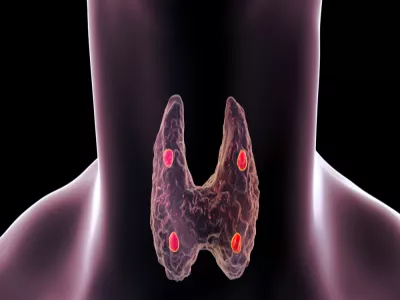
  • Hello world!

    Welcome to WordPress. This is your first post. Edit or delete it, then start writing!